

When it comes to diagnosing wrist injuries and conditions, X-ray imaging plays a crucial role in providing detailed insights into the underlying issues. Whether you have experienced a traumatic incident or are dealing with persistent wrist pain, a wrist X-ray can help healthcare providers determine the appropriate course of action.

Credit: www.mdpi.com
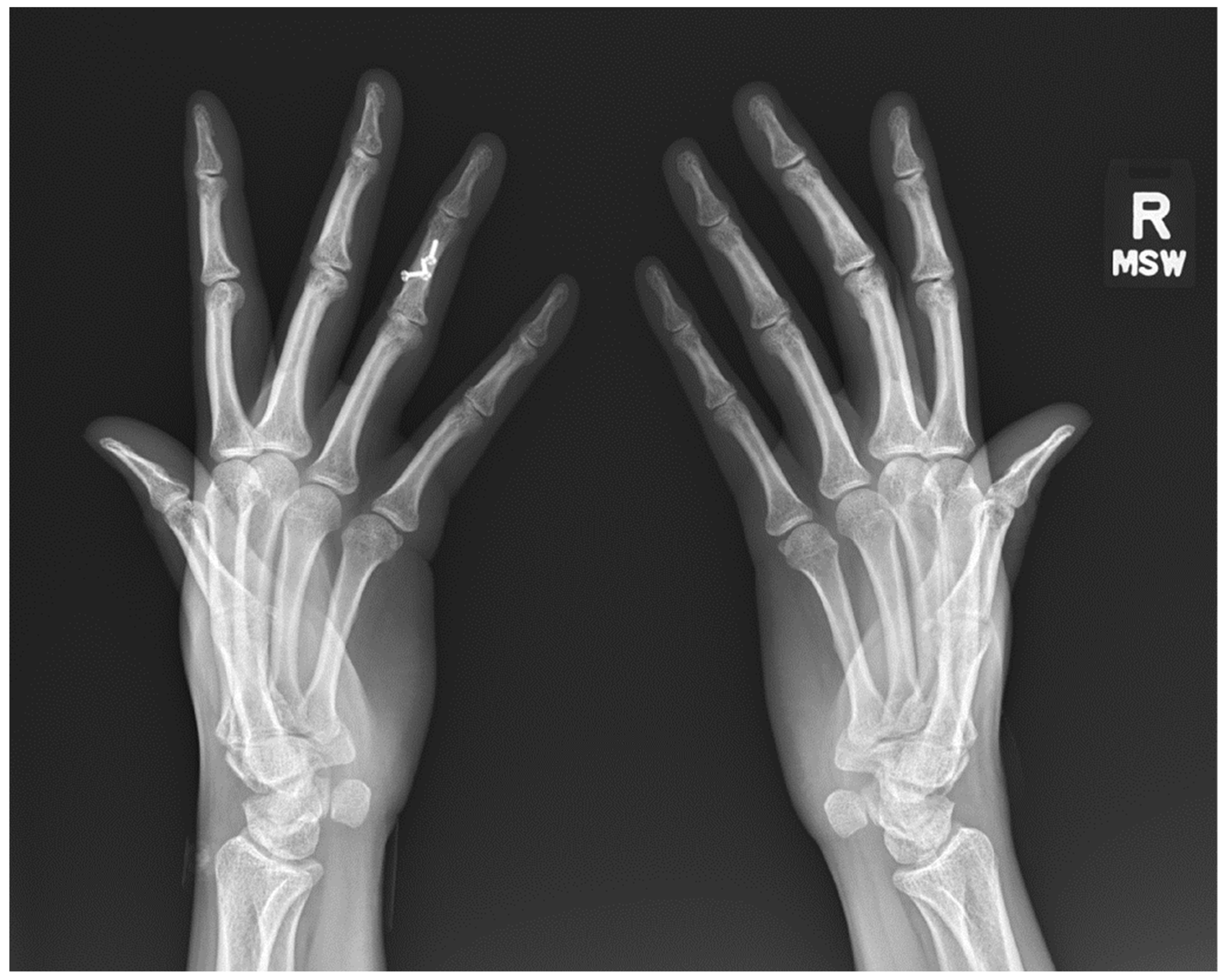
Understanding Wrist X-rays
A wrist X-ray is a quick, painless, and noninvasive procedure that involves capturing images of the bones in your wrist from different angles. The typical views include:
- Postero-anterior (PA) view
- Lateral view
- Oblique view
These views allow healthcare professionals to assess the alignment of the carpal bones, identify any fractures, and evaluate the overall condition of the wrist joint.
What Can A Wrist X-ray Reveal?
A standard wrist X-ray provides clear images of the two forearm bones – the radius and ulna – along with the eight wrist bones known as carpal bones. These images can help identify various conditions such as:
- Fractures
- Dislocations
- Arthritic changes
- Growth plate abnormalities
Common Reasons For Wrist X-rays
If you are experiencing wrist pain, swelling, tenderness, or deformities in the wrist joint, your healthcare provider may recommend a wrist X-ray to aid in diagnosis. Additionally, wrist X-rays are commonly used in cases of trauma or suspected fractures.
Credit: www.mdpi.com
Diagnostic Importance of Wrist X-rays
Accurate imaging techniques, such as X-rays, are crucial for evaluating the complex anatomy of the bones and soft tissues in the wrist. By examining these images, healthcare providers can make informed decisions regarding treatment options and further diagnostic procedures.
The Role Of Wrist X-rays In Trauma
In cases of wrist trauma, X-rays are often the initial imaging modality used to assess the extent of injuries. For instance, the scaphoid bone, a small bone in the wrist, is commonly evaluated for fractures following traumatic incidents.
Imaging Guidelines For Wrist Injuries
When a wrist injury occurs, healthcare professionals may recommend a series of X-ray views to thoroughly examine the affected area. These views can help identify specific issues and guide appropriate treatment plans.
Frequently Asked Questions Of Xray Wrist: Unlock The Secrets Of Your Wrist Health
What Is The Best Imaging For A Wrist Injury?
For a wrist injury, the best imaging options are MRI without contrast or ultrasound when the hand is displaced from its normal position. If there are penetrating injuries to the soft tissues of the hand or wrist that are not visible on x-ray, CT without contrast and ultrasound are appropriate.
The appropriateness criteria for acute hand and wrist trauma suggest MRI without contrast may also be appropriate.
What Are The Three Views Of The Wrist?
The wrist examination consists of three views: Postero-anterior (PA), lateral, and oblique. These views capture the full assessment of the wrist.
What Is A Xr Wrist 3 View?
A XR wrist 3 view is a diagnostic test capturing three angles: Postero-anterior (PA), Lateral, and Oblique views. It helps diagnose wrist conditions for patients with symptoms.
What Bones Are Visible In The Wrist?
The carpal bones, including the pisiform, are visible in the wrist X-ray images. The wrist X-ray shows the radius, ulna, and eight carpal bones in two rows.
Conclusion
Wrist X-rays are invaluable tools in the diagnosis and management of various wrist conditions and injuries. By providing detailed images of the wrist anatomy, X-ray imaging enables healthcare providers to deliver accurate diagnoses and personalized treatment plans for individuals seeking relief from wrist-related concerns.